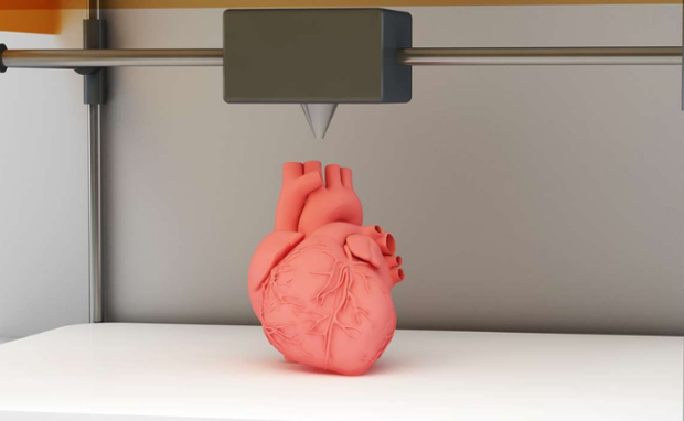
The Future of Cardiology: 3D Printed Heart Tissue
Medical technology has enabled doctors and researchers to create innovative solutions to the most pressing healthcare problems. One of them is using 3D printing to create organ transplants, a recent project from Stanford University scientists. It involves printing living tissue layer by layer until someone creates an entire organ.
Stanford researchers aim to create a 3D-printed human heart to revolutionize cardiology and organ transplantation. The process uses stem cells to create the desired organs, simplifying the organ creation process. As a result, it could save countless lives, especially those who need transplants.
This article will discuss the exciting Stanford project developing artificial transplant organs. I will explain how it works and how likely it is to launch in the future. Later, I will also cover the basics of 3D printing to further elaborate on the 3D printed tissue project. Whether you’re a medical expert or interested in medical innovations, this blog is for you.
How do you 3D print living tissue?
Mark Skylar-Scott, the assistant professor of bioengineering in the schools of Engineering and Medicine, is the head researcher behind 3D printing tissue. He explains how it works.
He and his team use advanced techniques to manufacture thick tissues one layer at a time. It places the exact types of cells needed at the correct spots.
It is like building a tower, brick by brick. Skylar-Scott noted it is suitable for replicating complex organs like the heart. Organs are complicated structures that must balance form and function.
How do Stanford scientists create 3D printer cells? They use stem cells, the ones the body uses to create specialized cells. Then, the researchers apply a special concoction to transform stem cells into organ cells for 3D printing tissue.
“Each line of stem cells we are developing are genetically engineered to respond to a specific drug,” he said. “Once they sense that drug, they differentiate into specific cell types.”
You may also like: ChatGPT Glasses Guides Users Through Interviews
Skylar-Scott’s technique is an example of bioprinting, which uses living cells to create organ-like structures. It is not a new concept, but it is usually slow. It prints one cell at a time. Consequently, printing a thousand per second will take over a thousand years to make a human heart.
However, the Stanford method sets itself apart by printing with cell clusters called organoids. “We take millions of those and condense them into what is essentially a human stem cell mayonnaise that we can then print through the printer,” Skylar-Scott said.
They take on the general shape of tissue that can have blood vessel networks printed within. The Stanford team printed a tube-like structure like a human vein that pumps fluid.
How does 3D printing work?
It is not easy to understand 3D printing tissue if you do not know how 3D printers work. I will elaborate on this process to get all readers up to speed.
Consumer-grade 3D printers create plastic components by stacking layers of molten resin. The machine feeds a spool of plastic filament into a nozzle.
The latter melts the plastic to let it pour into a surface. Then, it stacks liquid plastic layers to produce a specific shape. Nowadays, people use it to create custom-made toys.

Photo Credit: medicalbag.com
Others use this method to produce spare parts for broken devices. Also, some experts use 3D printing to make prototype medical devices and vehicle parts.
You may also like: Vietnam Successfully Clones Pigs
In contrast, 3D printing tissue is more complicated. It uses living cells, which are “soft, squishy, imperfect, and frustratingly fragile.” That is why creating an artificial heart could take thousands of years.
Professor Skylar-Scott admits he needs to scale up cell production so that 3D printing tissue becomes practical. However, it will require more than using a larger machine.
“We need to figure out ways to engineer cells to make them more robust and cheaper to grow, so we can start practicing and perfecting this method.” the Stanford professor said. “Once the pipeline for new cells is in place, I think we’re going to start seeing some incredible progress,” he added.
Conclusion
This 3D printing tissue project could revolutionize organ transplantation worldwide. If we could print organs, we could significantly reduce wait times for organ donations.
As a result, people could access life-saving treatments immediately. Professor Skylar-Scott and the other Stanford scientists need to develop it further, but it’s a step in the right direction.
As we wait for the project to be ready, everyone should stay up-to-date with the latest tips and trends. Fortunately, Inquirer Tech has you covered.
INQUIRER.net wants to hear from you! Take part in our reader survey and help us be better. Click on this image to answer.
Read Next
Subscribe to INQUIRER PLUS to get access to The Philippine Daily Inquirer & other 70+ titles, share up to 5 gadgets, listen to the news, download as early as 4am & share articles on social media. Call 896 6000.
For all the latest Technology News Click Here
For the latest news and updates, follow us on Google News.


