. DOI: 10.1117/1.NPh.10.2.025006")
Two-photon microscopy (TPM) has revolutionized the field of biology by enabling researchers to observe complex biological processes in living tissues at high resolution. In contrast to traditional fluorescence microscopy techniques, TPM makes use of low-energy photons to excite fluorescent molecules for observation. This, in turn, makes it possible to penetrate the tissue much more deeply, and ensures that the fluorescent molecules, or fluorophores, are not permanently damaged by the excitation laser.
However, some biological processes are simply too fast to be recorded, even with state-of-the-art TPMs. One of the design parameters that limits the performance of a TPM is the line scanning frequency, measured as frames per second (FPS). This refers to the rate at which the target sample can be swept across by the excitation laser along one direction (for instance, in a horizontal sweep). A slow scanning frequency also impacts the overall FPS of the system, since it determines how fast the laser can be swept across the other direction, i.e. in a vertical sweep. Together, these create a tradeoff between the microscope’s temporal resolution and the size of the observation frame.
Working around this problem, an international team of researchers from China and Germany recently developed a powerful TPM setup with an unprecedentedly high line scanning frequency. According to their report published in Neurophotonics, this microscopy system was designed for imaging fast biological processes at a high temporal as well as spatial resolution.
One of the key factors that distinguish the proposed TPM from the conventional ones is the use of acousto-optic deflectors (AODs) to control the scanning of the excitation laser. An AOD is a special type of crystal whose refractive index can be precisely controlled by acoustic waves. This, in turn, allows us to redirect a laser beam through it as desired. More importantly, AODs enable a faster laser-steering than that obtained with the galvanometers used in conventional TPMs.
Accordingly, the team designed a custom AOD with an exceptionally high acoustic velocity using a tellurium dioxide (TeO2) crystal, achieving a high line scanning frequency. With this AOD, the laser could scan a line in the frame within a mere 2.5 microseconds, corresponding to a maximum line scanning frequency of 400 kHz. Similarly, the team used an AOD to achieve a reasonable slow scanning frequency in the other direction.
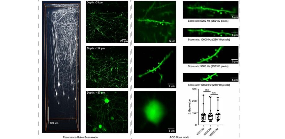
To further improve the adaptability of their microscope, the team added the option of switching to a galvanometer-based laser scanning mechanism when necessary. This allowed the scanning of large regions of the sample at an acceptable resolution and speed, making it easier to locate small areas of interest before switching to AOD scanning.
The team conducted several proof-of-concept experiments with the newly designed TPM. They installed cranial windows on genetically engineered mice and used them to observe the morphology and activity of neurons as well as the movement of single red blood cells (RBCs). The system achieved a frame rate of up to 10,000 FPS using an appropriate AOD configuration and frame size. This was sufficient to precisely measure the velocity at which calcium propagates in neuronal dendrites as well as to visualize the trajectory of individual RBCs within blood vessels.
Impressed by these results, Dr. Na Ji, Associate Editor of Neurophotonics and Luis Alvarez Memorial Chair in Experimental Physics at UC Berkeley, remarks, “The new system for AOD-based scanning microscopy represents a substantial improvement in imaging speed and performance, as demonstrated in its application for calcium signal propagation and blood flow measurements in the brain in vivo.”
Going forward, the new proof-of-concept TPM design will make it possible to capture fast biological processes, and could significantly improve our understanding of them.
More information:
Ruijie Li et al, Ten-kilohertz two-photon microscopy imaging of single-cell dendritic activity and hemodynamics in vivo, Neurophotonics (2023). DOI: 10.1117/1.NPh.10.2.025006
Citation:
New high-speed, two-photon microscope for precise biological imaging (2023, May 3)
retrieved 3 May 2023
from https://phys.org/news/2023-05-high-speed-two-photon-microscope-precise-biological.html
This document is subject to copyright. Apart from any fair dealing for the purpose of private study or research, no
part may be reproduced without the written permission. The content is provided for information purposes only.
For all the latest Science News Click Here
For the latest news and updates, follow us on Google News.