How microchip imaging cytometry makes laboratory testing more economical, easy-to-use, and accessible

A new publication from Opto-Electronic Advances addresses scientific and technical advances in the field of microchip imaging cytometry (MIC) and shows the applications of microchip imaging cytometry that may bring more economical, easy-to-use, and accessible healthcare to the public.
The economic globalization and the aging population of many countries in the world generate an enormous need for rapid and cost-effective point-of-need laboratory tests. Over the past two years, the entire world has been tackling challenges from COVID-19 pandemic. The general population in many countries are routinely taking nucleic acid tests and/or rapid antigen tests for screening purposes. Healthcare workers are in need of more economical, and easy-to-use diagnostic testing tools to support their healthcare practice. Public health agencies also need powerful diagnostic tools to help them make critical policy decisions.
In a typical clinic appointment, laboratory tests go through procedures such as lab requisition, sample collection, sample processing, and reporting. The average turn-around-time may vary from several hours to several days. For many disease diagnoses and monitoring requiring instant information and rapid decision-making, the traditional technology and workflow could not effectively meet the clinical needs.
Meanwhile, there is the “rapid test strip” option such as the COVID antigen test strip and the hCG pregnancy test strip that provides instant test results. These rapid test strips become an important diagnostic tool for screening and monitoring, although the application of the test strips is usually restricted to qualitative tests. Additionally, because of their relatively lower analytical sensitivity, these rapid test strips could not detect biomarkers that have a low quantity in the specimen. Therefore, there is a growing need to develop a quantitative, easy-to-use, and accessible diagnostic instrument and reagents.
Given the emerging healthcare needs, scientists and engineers continuously come up with creative diagnostic solutions using a variety of technological approaches. Among these technologies, microfluidics becomes a highly valuable approach to potentially address many of the requirements. Microchip imaging cytometry based on microfluidic technologies is such an innovative analytical platform that may change the landscape of the clinical lab testing field.
Microchip Imaging Cytometry (MIC) is a platform technology that can rapidly detect and analyze human biochemical substances such as cells, proteins, and nucleic acids. MIC devices have the attributes of portability, cost-effectiveness, and adaptability while providing quantitative measurements to meet the needs of laboratory testing in a variety of healthcare settings. Based on the use of microfluidic chips, MIC requires less sample and may complete sample preparation automatically. Therefore, they can provide quantitative testing results simply using a finger prick specimen. The decreased reagent consumption and reduced form factor also help improve the accessibility and affordability of healthcare services in remote and resource-limited settings.
The article reviews notable clinical applications of MIC technologies, such as HIV-patient monitoring, sickle disease screening, infectious disease diagnosis, etc. Depending on the level of automation and image capturing formats, MIC devices were classified into three approaches: Static-chip-static-fluid (SCSF), Static-chip-moving-fluid (SCMF) and Moving-chip-static-fluid (MCSF). Brightfield imaging, fluorescence imaging, and lens-free imaging techniques have been adopted in MIC systems. Image acquisition techniques such as time delay integration and temporally coded excitation were demonstrated to achieve higher sensitivity in detecting fast-moving objects in low light levels.
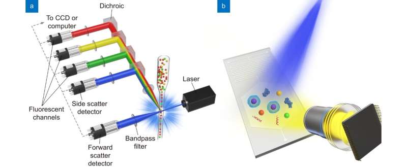
Compared with traditional flow cytometers, MIC analyzes objects such as cells and particles through a relatively wide and shallow microfluidic chip channel. As a result of the breakthrough development of semiconductor sensor devices and information technology in recent years, the light source and imaging detection components of MIC can also achieve higher optoelectronic performance.
Thanks to the innovation and development of biotechnology, micro-nano manufacturing, semiconductor materials, information technology, and other fields, MIC will find more important clinical test applications in the future, and promote the development of more economical, easy-to-use, and accessible point-of-need tests.
Recent advances in photonics, integrated optics, and imaging technologies promise to increase the sensitivity and functionality of MIC systems while decreasing their size and cost. Colors can be differentiated directly on the silicon CMOS image sensors using several techniques. Progress towards higher sensitivity detectors has also been made by integrating single-photon avalanche diodes in standard CMOS with microfluidic systems.
The development of MIC devices should focus on the following aspects: 1) the device should be portable to fit the diagnostic purpose in varying healthcare scenarios, 2) the device should be easy to use and provide sample-to-answer results rapidly (e.g. 15 minutes), 3) the microfluidic assembly should contain pre-loaded reagents and be disposable. Additionally, the analytical performance of MIC devices, such as sensitivity, accuracy, precision, robustness, needs to meet the certain testing requirements. In the process of instrument and reagent design and development, all these aspects need to be considered. Therefore, engineering design and development need to find the sophisticated balance between complexity, performance and cost, to meet the needs in healthcare and to benefit more patients.
Xilong Yuan et al, Microchip imaging cytometer: making healthcare available, accessible, and affordable, Opto-Electronic Advances (2022). DOI: 10.29026/oea.2022.210130
Provided by
Compuscript Ltd
Citation:
How microchip imaging cytometry makes laboratory testing more economical, easy-to-use, and accessible (2022, August 18)
retrieved 18 August 2022
from https://phys.org/news/2022-08-microchip-imaging-cytometry-laboratory-economical.html
This document is subject to copyright. Apart from any fair dealing for the purpose of private study or research, no
part may be reproduced without the written permission. The content is provided for information purposes only.
For all the latest Science News Click Here
For the latest news and updates, follow us on Google News.

