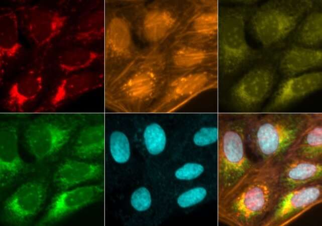
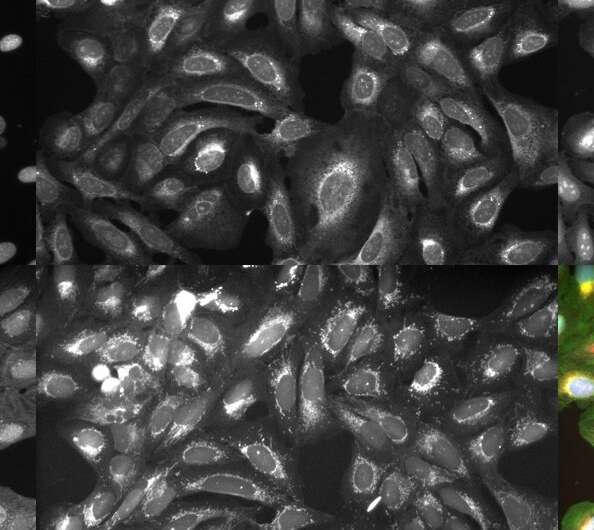
Comprehensive online guide makes quantitative bioimaging more accessible

Starting something new can be overwhelming to a person who does not know what they will need or where to start. Quantitative bioimaging, the process of obtaining quantitative data through images (sometimes thousands of them) and computational approaches, like machine learning, are relatively new to the field of microscopy.
Because of its novelty and complexity, biologists who do not have a computational background can have a difficult time incorporating quantitative bioimaging into their experiments. Resources do exist to teach everything needed to perform quantitative bioimaging, however, the volume of educational materials can intimidate those who are just getting started.
In collaboration with BioImaging North America (an organization that fosters collaboration in the bioimaging community), Beth Cimini, the associate director for bioimage analysis in the Imaging Platform at the Broad Institute of MIT and Harvard, has worked to make bioimaging accessible with the creation of bioimagingguide.org, an online guide for producing high quality microscopy images for quantitative data analysis.
With the guide and a companion paper published in PLOS Biology, Cimini, postdoctoral fellows Rebecca Senft and Barbara Diaz-Rohrer, and their colleagues aim to create a resource that makes the tools, techniques, and principles of quantitative bioimaging more accessible to biologists who don’t have a computational background.
We sat down with Cimini to discuss what bioimagingguide.org is and how it can help scientists who want to bring bioimaging into their research.
What is the main idea behind bioimagingguide.org?
There are many steps involved in creating good quantitative microscopy, and much to learn about each one of them. A lot of places run great, intensive microscopy courses, but those courses are held for around 20 people a year. And maybe you’re lucky enough to work at a place with a great microscopy center where they run a course like this every year. A lot of people aren’t, however.
What we felt was missing was a “big picture” overview of microscopy that was not a full textbook, but a mid-sized guide that says, “here are all the things you have to learn and here are links to great resources that we or other people have written to learn more.”
So our goal was to put together something that provides a comprehensive outline of the topics one needs to know to get started in bioimaging, and which links people to information that they can reference at their own pace.

The guide originally started as just a paper, but we soon realized that there was too much information. Also, we wanted a way to add new things as we find them and keep the guide up to date. A paper is a static point in time and shows what information was available when it was released; if somebody writes a great new thing tomorrow, we wanted to be able to bring it in.
What types of bioimaging experiments did you have in mind while writing the guide?
Bioimaging experiments can range from a single image where someone wants to measure one thing to terabytes of images where they want to measure thousands. A lot of what we cover in the guide is aimed at the person who needs to stain a sample, take a few dozen fluorescent images, and learn two or three things from those images.
How is this guide different from other resources?
This guide is unique in that it teaches the major topics of bioimaging to beginners in an accessible, online format that brings together and organizes a great deal of content that already existed elsewhere. We intentionally structured it so that people could ingest information at a broad level, but if they want details, they can easily find more information. Not everything is presented on the screen at once, which I hope can make it less overwhelming to start. Rebecca and Barbara did a tremendous job leading roughly 15 authors on the paper and putting together some great resources.
What inspired you all to create the guide?
In the Imaging Platform, we make open-source tools and workflows for researchers at Broad and beyond. On average, we collaborate with 20 to 30 groups a year on projects, and talk to hundreds of researchers. In talking to these folks, we often hear the same frustrations about having to repeat parts of a project because of errors that could have been avoided if they had known what to do.
We had already wanted to put together a way to help people generate good data suitable for good quantification, and in these conversations saw a need for an introduction on how to perform good microscopy as well. So we reached out to BioImaging North America’s Image Informatics Working Group , which works on image analysis, and Training and Education Working Group, which works on microscopy education, and asked for their advice on creating a project like this.
![A bundle of mouse trophoblast stem cells imaged using fluorescent microscopy. Credit: Rivron et al. 2018, available from the Broad Bioimage Benchmark Collection [Ljosa et al., Nature Methods, 2012] A how-to for performing quantitative bioimaging](https://scx1.b-cdn.net/csz/news/800a/2023/a-how-to-for-performin-2.jpg)
What is your favorite feature on the website?
There are a lot of features to love, but the fact that almost all the information in the guide searchable first comes to mind. There are many links to papers and other websites, and also a glossary full of terms and definitions.
I’m also really excited about a feature that we’re working on, but which isn’t live yet: automatic translation of the website. Not every scientist is fluent in English, and while someone can always use Google Translate or something like that, such translations are not always perfect. We are working with the Training and Education Working Group to translate all of the content into French, Spanish, and Portuguese, and are collaborating with people to get the site translated into other languages as well.
How do you expect or hope that people will use this guide?
Very often you’ll find quantitative bioimaging does not happen linearly, but rather in a circle. A person might get to a point in their project and realize that something that they did previously has made it so that they can’t proceed. They must then go back to the bench and try a different antibody or dye, or even a different kind of microscope.
My hope is that the information we’ve compiled will help people recognize where they are on that circle and get around it faster to the answers they seek.
More information:
Rebecca A. Senft et al, A biologist’s guide to planning and performing quantitative bioimaging experiments, PLOS Biology (2023). DOI: 10.1371/journal.pbio.3002167
Citation:
Comprehensive online guide makes quantitative bioimaging more accessible (2023, July 14)
retrieved 15 July 2023
from https://phys.org/news/2023-07-comprehensive-online-quantitative-bioimaging-accessible.html
This document is subject to copyright. Apart from any fair dealing for the purpose of private study or research, no
part may be reproduced without the written permission. The content is provided for information purposes only.
For all the latest Science News Click Here
For the latest news and updates, follow us on Google News.

