An interdisciplinary solution for enhanced high-resolution imaging in electron and optical microscopy

Although electron microscopy can already reveal details as small as one nanometer, ongoing research seeks to break through barriers limiting image quality and reducing the optical dose on the samples. Aberration is a common problem in electron microscopy that can reduce the resolution and quality of the images produced.
Additional complex phase and amplitude controls are needed in these microscopes. An international team of researchers led by Akhil Kallepalli (Kallepalli Lab) working within the Optics Group at the University of Glasgow set out to address the problem. Working from an optics perspective, they developed and tested a new ghost imaging algorithm, finding that they could produce an image with improved resolution and contrast using lower flux illumination, which could reduce sample damage.
The research was published Dec. 21 in Intelligent Computing.
Optical modulation is required to achieve better control of illumination strategies. Modulation in optics is the process of varying the properties of light waves to encode information. It is used in optical communication systems and in various applications, such as spectroscopy and imaging. Modulators of many kinds have long been available in the field of optics.
However, modulators are not available for electron microscopy. It is still challenging to achieve complex phase and amplitude control to reduce phase aberration for continuous imaging enhancement in the field of electron microscopy.
The authors applied computational ghost imaging, an optical approach, to electron microscopy and designed a new algorithm to address this problem. The approach inverts knowledge of the projected patterns and their measured transmission to reconstruct the image. This can be used to measure the transmittance of the sample when illuminated with more complex spatial patterns.
In this system, the resulting form of the light field in the plane of the object can be calculated using numerical beam propagation techniques enabling both lens-free and far-field implementations. Therefore, computational ghost imaging can be used for transmission electron microscopy imaging.
In optical methods, spatial light modulators can be used to ensure the orthogonality of the imaging patterns. It is, however, difficult to guarantee orthogonality between patterns when using natural scattering or highly limited modulators. This new algorithm designed by the authors makes optimal use of patterns regardless of their orthogonality. They call their new method “orthogonalized ghost imaging.”
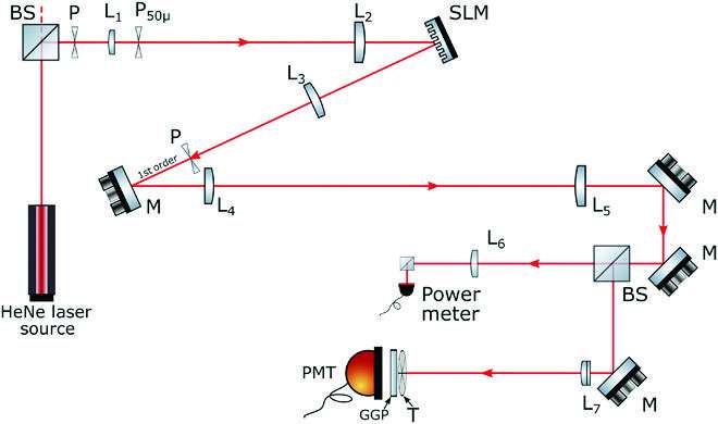
The authors tested their method in two ways. First, they conducted an optical experiment analogous to the transmission electron microscopy system. This experiment tested the illumination strategy and the algorithm’s robustness to non-orthogonality. After that, they tested their method with transmission electron microscopy.
The experiments showed that the authors’ ghost imaging algorithm produces a higher-resolution image reconstruction with better contrast in comparison to the most common online ghost imaging algorithm. The new algorithm enhances the imaging capabilities at any wavelength, and is robust to the non-orthogonality of the pattern sets, enabling effective application in both optical and electron microscopy.
In an appendix to their paper, the authors highlight some findings relating to electron microscope sample damage, which could be reduced using their method. Future development can be used to further optimize imaging resolution or speed across both optical and electron microscopic imaging.
More information:
Akhil Kallepalli et al, Challenging Point Scanning across Electron Microscopy and Optical Imaging using Computational Imaging, Intelligent Computing (2022). DOI: 10.34133/icomputing.0001
Provided by
Intelligent Computing
Citation:
An interdisciplinary solution for enhanced high-resolution imaging in electron and optical microscopy (2023, March 3)
retrieved 3 March 2023
from https://phys.org/news/2023-03-interdisciplinary-solution-high-resolution-imaging-electron.html
This document is subject to copyright. Apart from any fair dealing for the purpose of private study or research, no
part may be reproduced without the written permission. The content is provided for information purposes only.
For all the latest Science News Click Here
For the latest news and updates, follow us on Google News.

