
A team of researchers at the University of California at Berkeley, working with a group at Lawrence Berkeley National Laboratory, has developed a non-invasive way to image Wigner crystals directly. In their paper published in the journal Nature, the group describes their approach and explain how it could be used to advance research regarding Wigner crystal states. Carmen Rubio-Verdú with Columbia University has published a News & Views piece outlining the nature of Wigner crystals and describing the work by the team in the same journal issue.
Wigner crystals have a crystal lattice structure that forms when electrons are sparsely spaced in certain 2D materials. They have been observed in materials such as 2D semiconductors and liquid helium, but they are notoriously difficult to observe or image because they are so fragile. In this new effort, the researchers have developed a way to view Wigner crystals without disturbing them, which allows for more precise imaging.
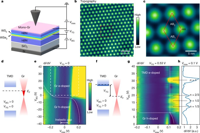
The researchers placed a thin sheet of tungsten disulfide on top of a thin sheet of tungsten diselenide, creating a tiny heterostructure. Notably, both are transition-metal dichalcogenides, and in this instance, were just 1 nanometer thick. The team then added electrons to both layers, which naturally formed into 2D structures, though the spacing between the electrons was slightly smaller in one of the layers. The mismatch in the electron patterns resulted in the creation of a moiré pattern which was also a Wigner crystal. The researchers then placed a layer of graphene on top of their heterostructure to protect the crystal structure below. Then they used a scanning-tunneling microscope to create images of the crystals without disturbing them. Later, the team added a layer of hexagonal boron nitride to the heterostructure to better protect it, allowing probing with the microscope.
The researchers also tried adding and removing electrons from the structure prior to adding the protective barriers and found that doing so resulted in the crystal structures forming into shapes including triangles or hexagons. Rubio-Verdú suggests the new technique could lead to new methods to image other tiny, fragile structures.
Researchers trap electrons to create elusive crystal
Hongyuan Li et al, Imaging two-dimensional generalized Wigner crystals, Nature (2021). DOI: 10.1038/s41586-021-03874-9
Carmen Rubio-Verdú, Electron crystals come under the microscope, Nature (2021). DOI: 10.1038/d41586-021-02573-9
© 2021 Science X Network
Citation:
A non-invasive way to image Wigner crystals directly (2021, October 1)
retrieved 1 October 2021
from https://phys.org/news/2021-10-non-invasive-image-wigner-crystals.html
This document is subject to copyright. Apart from any fair dealing for the purpose of private study or research, no
part may be reproduced without the written permission. The content is provided for information purposes only.
For all the latest Science News Click Here
For the latest news and updates, follow us on Google News.